Cellular 2-photon imaging
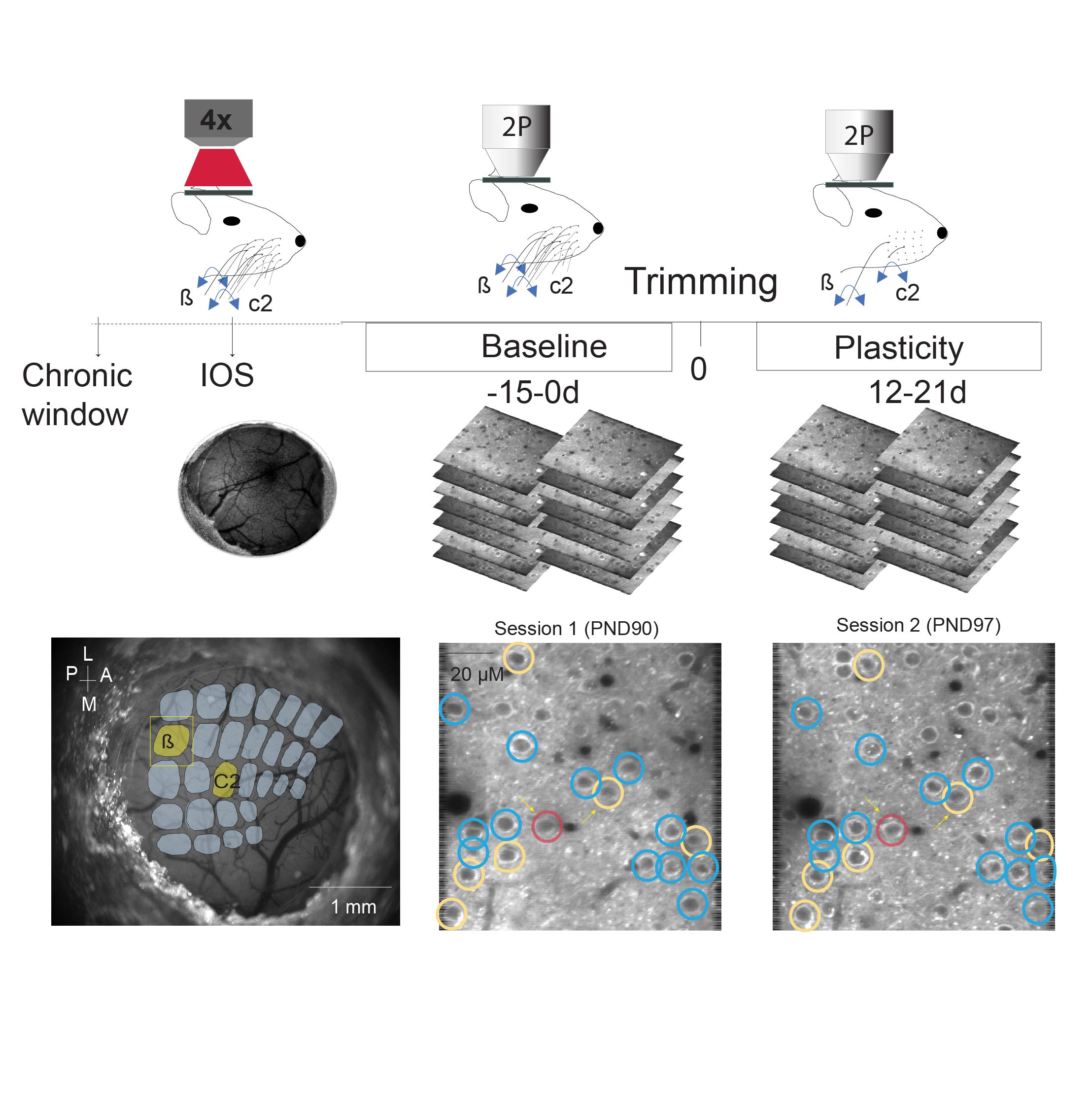
Experience dependent changes in wS1 L2/3 population activity in Syngap1 mice. (A) Experimental timeline. β and C2 whiskers were stimulated in two two-photon imaging sessions (10 deflections at 20 Hz). Then, whiskers were trimmed (all but β) every two days and animals were imaged two more sessions (β and C2 whisker stimulation, 10 deflections at 20 Hz). (B) Image of cortical surface through a chronic cranial window, imaging areas indicated in yellow (β and C2). Representative in vivo two-photon microscopy images (top).
Axonal 2-photon imaging
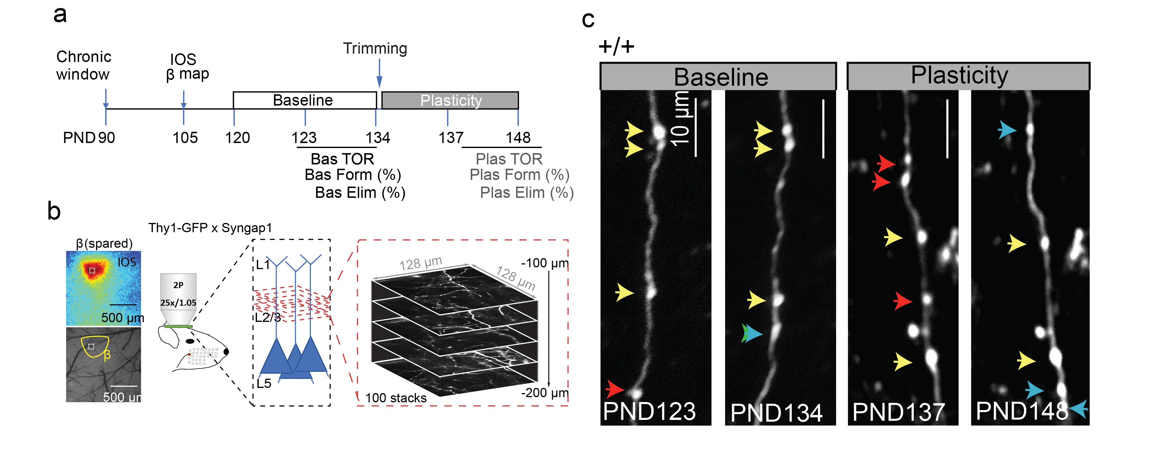
Experience-dependent reorganization of synaptic inputs in wS1 in models of neurodevelopomental disordersv. (A) Experimental timeline. Thy1-GFP x Syngap1 mice were implanted with a chronic cranial window and recovered for at least two weeks before Intrinsic Optical Signal (IOS) imaging. IOS was used to identify the location activated by the deflection of β whisker. After three baseline two-photon sessions were performed, whiskers were trimmed (all but β) every two days and animals were imaged two more sessions. (B) Representation of in vivo imaging procedure of axonal bouton segments in wS1 L2/3 in Thy1-GFP x Syngap1 mice. (C) Images obtained in different baseline and plasticity sessions showing axonal dynamics in WT mice. Yellow arrows indicate stable axonal boutons, blue arrows indicate new boutons and red arrows indicate lost boutons.